
AI Professor Courses
How Can I Use AI-Driven Simulation to Visualize and Master Complex Histology in Medical Studies?
Mastering the microscopic structures of the human body is essential for medical students. Learn how AI-driven simulation and targeted tutoring can simplify complex histology, allowing for faster memorization and deeper clinical understanding through interactive 24/7 learning.
Direct answer: You can master complex histology by using AI-driven simulation to generate interactive, annotated virtual slides that highlight specific cellular structures in real time. AI Professor Courses offers over 80 courses across AI, Sciences, Math, Nursing, Law, History and more, starting at just $5/month, providing 24/7 instant tutoring to guide your medical visualization studies.
How does AI-driven simulation bridge the gap between static diagrams and microscopic reality?
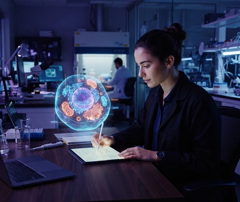
AI-driven simulation bridges the gap by transforming two-dimensional, static images found in traditional textbooks into interactive, multi-dimensional models that respond to your queries. While traditional histology involves peering through a microscope at slides that may be faded or poorly stained, AI tools can synthesize ideal representations of tissue samples, allowing you to manipulate focus, staining intensity, and magnification levels instantaneously. This provides a level of clarity that physical lab settings often struggle to maintain due to sample degradation or limited access to high-end equipment.
By integrating these simulations into your study routine, you move from passive observation to active engagement. Instead of just memorizing the appearance of a simple squamous epithelium, you can ask an AI to simulate the tissue under various pathological states, such as inflammation or fibrosis. This teaches you not just what healthy tissue looks like, but how physiological changes alter its microscopic architecture. This iterative process of visual hypothesis testing is fundamental to clinical diagnostic skills. When you understand the biological processes causing visual changes in a tissue sample, you move beyond rote memorization into the realm of conceptual synthesis, which is the hallmark of advanced medical proficiency.
Furthermore, the ability to layer AI-generated annotations onto these simulations allows for a guided discovery approach. You can toggle labels for organelles, cellular junctions, and extracellular matrix components, effectively creating a personalized anatomical atlas. When you reach a point of confusion—perhaps distinguishing between two similar types of glandular tissue—the AI can generate comparative side-by-side simulations that specifically emphasize the subtle differences in nuclear density or cytoplasmic granular distribution. This dynamic feedback loop ensures that your study sessions are always calibrated to your current knowledge level, preventing the frustration of wading through irrelevant material while ensuring you do not skip over foundational concepts that are vital for your upcoming examinations.
Why is 24/7 AI tutoring superior to traditional study groups for histology?
Traditional study groups often suffer from scheduling conflicts and varying levels of understanding among participants, whereas 24/7 AI tutoring provides consistent, expert-level clarity whenever you need it. Histology is a subject that requires frequent, short bursts of review rather than long, infrequent cramming sessions. With AI Professor Courses, you can access instant tutoring at 3:00 AM after a late-night lab session, ensuring that you resolve misunderstandings regarding epithelial arrangements or connective tissue fibers before they become entrenched as incorrect knowledge. The friction of waiting for a professor's office hours or waiting for a study group to convene can lead to knowledge gaps that compound over the duration of a medical program.
Beyond simple availability, AI tutoring offers a private, non-judgmental environment where you can ask fundamental questions repeatedly until they are mastered. In many educational settings, students may feel pressured to project confidence, which hinders their ability to voice genuine confusion. An AI tutor, by contrast, functions as a tireless guide that adapts to your learning pace. If you need to spend an hour focusing on the microscopic differences between various types of cartilage, the AI will remain patient, providing as many examples, simulations, and Socratic prompts as necessary. This consistency builds a stronger, more resilient foundation for your medical career, where precise diagnostic skills are non-negotiable.
Consider the following advantages of adopting an AI-centered approach for your medical studies:
- Personalized pacing that prevents burnout while maintaining academic rigor.
- Immediate access to complex histological simulations without the need for physical slide preparation.
- Ability to pivot across disciplines instantly, including our comprehensive modules in:
- Sciences and Nursing
- Anatomy and Physiology
- Molecular Biology and Genetics
- Clinical Diagnostics and Pathology
- Reduction of memory load through AI-driven active recall and spaced repetition algorithms.
Can AI tools help you transition from pattern recognition to clinical reasoning?
AI tools facilitate the transition from pattern recognition to clinical reasoning by linking histological findings directly to systemic disease processes and patient symptoms. While recognizing a specific tissue type is a critical first step, the ultimate goal of medical study is to understand the clinical significance of that finding. AI simulations can provide a clinical vignette alongside the histology, prompting you to reason through how a particular structural abnormality discovered under a microscope would manifest as a physiological deficit in a patient. This contextual learning is significantly more effective than isolated visual memorization because it embeds information into a clinical framework.
For example, if you are studying the histology of renal glomeruli, the AI can present you with a simulated histological slide showing damage to the basement membrane. Simultaneously, it can prompt you to analyze the potential impact on glomerular filtration and patient urinalysis results. This forces you to connect microscopic morphology with macroscopic health outcomes. This method effectively mimics the cognitive process of a pathologist, who must interpret visual data in the context of a patient's history and symptoms. By practicing this linkage repeatedly, you develop a mental map that links the cellular level of existence to the whole-body clinical presentation, which is essential for success in medical boards and subsequent clinical practice.
Moreover, the AI can track your reasoning process, providing feedback when your logic deviates from established clinical pathways. If you misinterpret the relationship between specific collagen distribution in a tissue and the tissue's tensile strength, the AI will immediately identify the error and offer a corrective simulation or explanation. This immediate feedback loop is critical for correcting biases in your reasoning early in your education. As you accumulate more clinical knowledge, the AI can increase the complexity of these vignettes, ensuring that your training remains challenging yet manageable. By the time you reach clinical rotations, you will have moved well beyond merely naming cell types; you will be prepared to evaluate complex tissue samples as part of a comprehensive diagnostic process, making your clinical interactions significantly more productive.