AI Professor Courses
How Can I Effectively Use AI-Driven Simulation to Visualize and Master Complex Histology in Medical Studies?
Learn how to use AI-powered simulation tools to master complex histological structures, identify microscopic tissues, and improve clinical diagnostic skills in medical studies.
Direct answer: You can master complex histology by using AI-driven simulation to generate high-resolution, interactive 3D visualizations of tissue structures, which bridges the gap between static two-dimensional microscope slides and living anatomy. AI Professor Courses supports your journey with over 80 courses across Sciences, Nursing, and Medicine, offering 24/7 personalized tutoring for just $5/month to help you excel.
How does AI bridge the gap between static slides and real tissue?
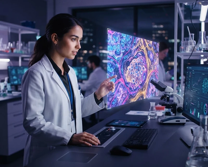
Traditional histology training relies heavily on static, often faded, glass slides that fail to represent the dynamic, three-dimensional reality of human physiology. AI bridges this divide by synthesizing vast datasets of high-resolution images to create interactive, rotatable, and zoomable simulations of cellular architecture. Instead of spending hours squinting at a two-dimensional thin section of liver tissue, an AI tutor can walk you through the structural hierarchy of a hepatic lobule, demonstrating exactly how the portal triad functions within the three-dimensional context of the organ. This transition from passive observation to active exploration is the hallmark of modern medical pedagogy.
Furthermore, AI-driven platforms can normalize the visual data you see. In standard laboratory environments, staining quality varies wildly based on lab techniques, reagents, and slide age. AI simulation models standardize these images to highlight critical diagnostic markers, such as specific intracellular organelles or extracellular matrix proteins. By reducing the noise inherent in suboptimal lab samples, AI allows you to focus your mental energy on pattern recognition—identifying the difference between a malignant growth and healthy tissue morphology—rather than struggling with the limitations of physical equipment. This is particularly vital for medical students in their early clinical years who are attempting to build a reliable mental library of what 'normal' versus 'pathological' looks like under the lens.
Finally, the integration of AI allows for adaptive difficulty levels in your histology practice. An AI tutor can recognize when you are consistently misidentifying specific tissue types, such as the subtle differences between pseudostratified columnar epithelium and simple columnar epithelium. It will then adjust the simulation parameters to present you with comparative visual challenges, forcing you to articulate the morphological distinctions. By engaging in this iterative feedback loop, you move past mere memorization of textbook diagrams and develop an intuitive, expert-level grasp of cellular structure that is both faster to build and more resistant to forgetting over the long term.
How can I use AI to simulate diagnostic workflows?
AI-driven simulations allow you to replicate the actual clinical workflow of a pathologist, enabling you to practice diagnostic reasoning in a risk-free environment where every error serves as a teaching moment. Rather than just identifying tissues in isolation, you can engage with simulated 'patient cases' where the AI presents you with a clinical history followed by a series of virtual biopsy samples. You are then tasked with formulating a diagnosis, justifying your reasoning based on the histological markers visible in the simulation, and receiving immediate, evidence-based critique from the AI tutor. This process forces you to integrate your histological knowledge into the broader clinical picture, which is essential for success in both board examinations and real-world medical practice.
This workflow-based training is far superior to rote memorization because it fosters the development of clinical 'pattern recognition,' a skill that typically takes years to cultivate. When you interact with AI simulations, you are not just seeing a slide; you are seeing the culmination of symptoms, patient age, and tissue characteristics. For example, if you are studying renal pathology, the AI can present you with a series of glomerular structures ranging from healthy to various stages of nephropathy. By simulating the progression of disease, the AI helps you internalize how specific physiological processes manifest physically at the cellular level. This is the cornerstone of clinical mastery, ensuring that when you finally interact with actual patient data, your brain has already built the necessary neural pathways to interpret what you are seeing.
Additionally, the diagnostic framework provided by AI platforms offers an objective standard for success. In a traditional setting, your understanding is limited by the subjective feedback of a human professor who may be pressed for time. AI-driven platforms evaluate your performance based on a massive database of diagnostic standards. They can highlight exactly which histological feature you missed—perhaps a specific basement membrane thickening or a subtle infiltration of inflammatory cells—and link that finding directly to the underlying pathology. This precision is unparalleled in traditional learning environments and allows you to calibrate your diagnostic eye to an exact, repeatable standard, minimizing the uncertainty that often plagues students in their first clinical rotations.
Why is active retrieval with AI better than passive atlas review?
Active retrieval, the psychological process of pulling information from your brain rather than feeding it in, is the most effective way to solidify knowledge, and AI provides the perfect scaffolding for this effort. While an atlas or a textbook provides a static reference that invites passive scanning, an AI tutor creates a 'testing' environment. By using AI to hide labels, challenge you to identify tissues from randomized angles, or present variations of common structures, you force your brain to perform the heavy lifting of recall. This effortful processing, while initially more mentally taxing than reading an atlas, leads to significantly higher retention rates, ensuring that the complex terminology of histology becomes second nature rather than a list of terms you only recognize on an exam.
This method of study is applicable across many disciplines, though the depth required for medical studies is particularly high. To see the breadth of how our systems support your learning, consider these core subject categories available through AI Professor Courses:
- Medical and Nursing (Histology, Anatomy, Pathology, Pharmacology)
- Core Sciences (Biology, Chemistry, Physics)
- Mathematics (Calculus, Statistics, Linear Algebra)
- Professional Fields (Law, Criminal Justice, History)
By focusing on these areas, we ensure that no matter your academic path, you have the tools to practice active retrieval. When you use AI to test your knowledge of histology, you are essentially engaging in a form of spaced repetition. The AI tracks the specific tissues or structures you struggle to classify and systematically reintroduces them in future sessions. This prevents the common student trap of 'familiarity,' where you mistakenly believe you know a concept because you have seen the image multiple times. By forcing you to generate the correct identification from scratch, the AI ensures you are actually mastering the material. This distinction is critical for medical students, where deep mastery of microscopic anatomy is a requirement for safe clinical practice. By moving from passive viewing to active, AI-guided interrogation, you turn every study hour into a high-yield investment, setting yourself apart from those relying on traditional, less efficient study habits.